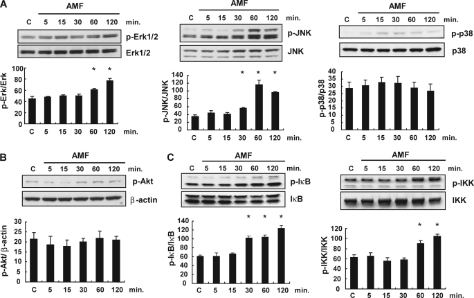
FIGURE 4.
Basal and activation statuses of the MAPK, phosphatidylinositol 3-kinase, and NFκB pathways in SBcl-2 cells treated with AMF. SBcl-2 cells were stimulated with rAMF (500 ng/ml), and the cells were extracted after 5, 15, 30, 60, and 120 min. The levels of phosphorylated and total proteins were detected by Western blotting analysis. C, control samples. A, time-dependent changes in the expression levels of MAPKs, phospho-ERK (p-Erk), ERK, phospho-JNK (p-JNK), JNK, phospho-p38, and p38, in response to rAMF stimulation. Densitometric analyses of the data are shown under the Western blotting bands. *, p < 0.05 versus control cells. B, time-dependent changes in the expression levels of phospho-Akt and β-actin in response to rAMF stimulation. C, time-dependent changes in the expression levels of the NFκB pathway, phospho-IκB, IκB, phospho-IKK, and IKK, in response to rAMF stimulation. Densitometric analyses of the data are shown under the Western blotting bands. *, p < 0.05 versus control cells. Data represent the means ± S.E. of triplicate analyses.

