FIGURE 2.
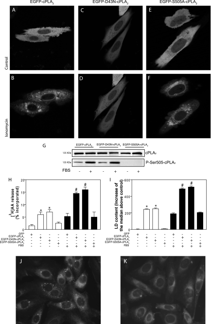
EGFP-D43N-cPLA2α is as effective as EGFP-cPLA2α to induce LD biogenesis in response to FBS. A–F, CHO-K1 cells were transiently transfected with plasmids encoding EGFP-cPLA2α (A and B), EGFP-D43N-cPLA2α (C and D), or EGFP-S505A-cPLA2α (E and F). Confocal images were obtained before (A, C, and E) or 10 min after addition of 5 μm ionomycin. G, Western blots of total cPLA2α and phospho-Ser-505-cPLA2α of transiently transfected cells. H, [3H]AA release from serum-starved CHO-K1 cells transfected with EGFP-cPLA2α, EGFP-D43N-cPLA2α, or EGFP-S505A-PLA2α and kept unstimulated (open bars) or stimulated with 7.5% FBS for 15 min (filled bars). I, indirect quantification of LD by flow cytometry in cells after 6 h under control conditions (open bars) or with 7.5% FBS (filled bars). J and K, serum-starved CHO-K1 cells transiently transfected with EGFP-D43N-cPLA2α (J) or with EGFP-S505A-PLA2α (K) were stained with Nile red for epifluorescence microscopy. Results in H and I are means ± S.E. of three independent experiments. *, significantly different (p < 0.01) from green fluorescent protein-transfected cells; #, significantly different (p < 0.01) from FBS-stimulated, green fluorescent protein-transfected cells.

