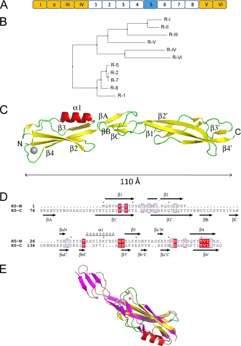
FIGURE 1.
X-ray crystal structure of the mucus-binding protein Mub-R5 repeat. A, the organization of the modular repeat region of MUB from L. reuteri 1063. Repeats are labeled according to the nomenclature described in (Ref. 23). Type 1 repeats (RI to RVI) are colored gold and type 2 white except for the R5 repeat, which is highlighted in blue. B, neighbor joining tree as calculated by JALVIEW (82) for the non-redundant set of repeat sequences based on the percentage identity at each aligned position (note that repeats R2, R4, and R6 are identical as are R3 and R5). C, architecture of Mub-R5. α-Helices are colored red, and β-strands are yellow. The N and C termini of the protein are labeled, as are the major secondary structural elements. The single calcium ion bound to the N-terminal domain is represented as a gray sphere. D, structure-based sequence alignment of the B1 (R5-N) and B2 (R5-C) domains of Mub-R5. Secondary structural elements in both proteins are indicated and labeled. Identical residues are highlighted in red. E, an overlay of the structures of the B1 and B2 domains. The secondary structural elements of the B1 domain are shown in red (α-helices) and yellow (β-strands) and in magenta (β-strands) for the C-terminal B2 domain.

