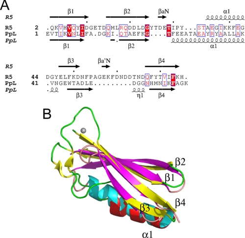
FIGURE 2.
Structural similarity between the B1 domains of Mub-R5 and Protein L. A, a structure-based alignment of the sequences of the B1 domain of Mub-R5 (R5) and the B1 domain of Protein L (PpL). Secondary structural elements in both proteins are indicated and labeled. Identical residues are highlighted in red. A single turn of a 310 helix labeled η1 preceding strand β4 in the structure of Protein L is absent from the R5 domain. B, an overlay of the structures of the B1 domains of Mub-R5 and PpL. The secondary structure of the Mub-R5 B1 domain is shown in red (α-helices) and yellow (β-strands) with the coil in green. For Protein L, α-helices are colored cyan and β-strands are magenta with coil regions in pink. The calcium ion bound to the Mub-R5 B1 domain is shown as a gray sphere.

