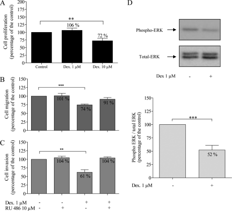
FIGURE 1.
Effects of dexamethasone (Dex) (1 and 10 μm) and RU 486 (10 μm) on U373 MG cells. Cell proliferation (A, n = 15) was evaluated after 48 h using the MTT assay. Migration (B, n = 7) and invasion (C, n = 5) were assessed using the modified Boyden chamber assays as described under “Experimental Procedures.” Representative Western blot of phospho-ERK1/2 and total ERK1/2 (D) and quantification of Western blot analysis (ratio of phospho-ERK1/2/total ERK1/2) (n = 9). *, p < 0.05; **, p < 0.01; ***, p < 0.005. Results are expressed as mean ± S.E. (error bars).

