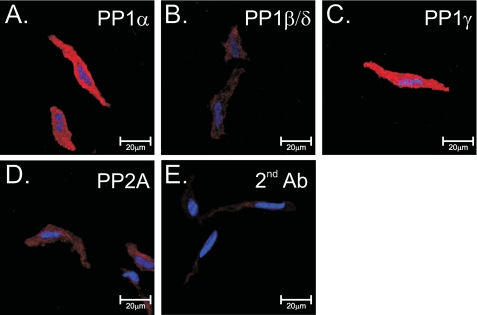
FIGURE 2.
Immunodetection of protein phosphatases in rabbit pulmonary artery smooth muscle cells. Shown is immunolabeling of isolated cells for PP1α (A), PP1β/δ (B), PP1γ (C), and PP2A (D), all detected using a biotinylated anti-goat antibody (red) with streptavidin. E, cells labeled with secondary antibody only. For all images, nuclei were stained with bisbenzamide (blue). Image series were taken in the z-dimension at 0.5-μm intervals, and a full cross-section of the cell was selected for display purposes.

