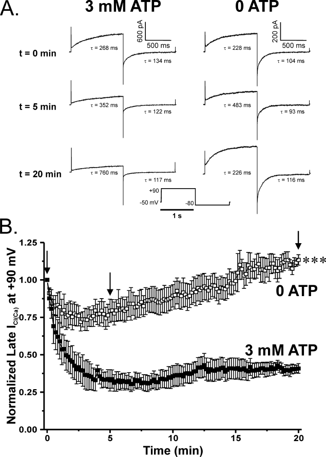
FIGURE 3.
Effects of altering ATP levels and the state of global phosphorylation on Ca2+-activated Cl− currents elicited by 500 nm Ca2+. A, representative current traces evoked with a 500 nm Ca2+ pipette solution containing either no ATP or supplemented with 3 mm ATP. Under conditions supporting global phosphorylation, both the time-dependent outward current at +90 mV and the inward tail current at −80 mV were substantially attenuated over the course of 20 min. However, PA myocytes dialyzed in the absence of exogenous ATP showed enhanced ICl(Ca), following a transient decline in amplitude, which continued to grow beyond the initial current over the course of the 20-min protocol. Time constants (τ) of activation and deactivation are shown for each representative trace and are displayed for both sets of experiments at t = 0, 5, and 20 min. B, plot showing the mean time course of changes of normalized late current at +90 mV (1-s steps at 10-s intervals). Currents were normalized against the initial current recorded immediately following membrane rupture (t = 0) and plotted as a function of time. Cells dialyzed with 3 mm ATP (filled squares; n = 4) displayed ICl(Ca) that declined to ∼40% of the initial level after 20 min. ICl(Ca) recorded from cells dialyzed with a solution lacking ATP (empty squares; n = 7) ran down to about 75% of its initial amplitude. Recovery of ICl(Ca) in these cells could be seen as early as 3 min to equal initial amplitude after 15 min and finally exceed it by ∼10% after 20 min. ***, significantly different from 3 mm ATP with p < 0.001.

