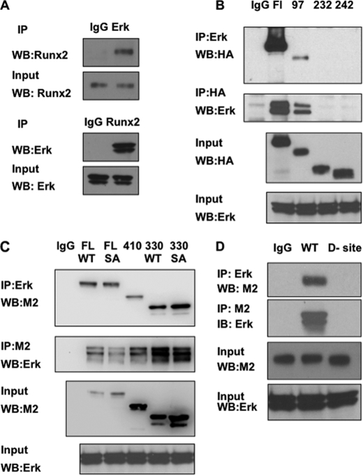
FIGURE 4.
Association of Runx2 with ERK. A, co-immunoprecipitation of endogenous Runx2 and ERK. MC-4 cell nuclear extracts were immunoprecipitated with IgG, ERK, or Runx2 antibodies and blots were probed as indicated. B–D, identification of the ERK binding domain in Runx2. Wild type (WT) Runx2 or the indicated N-terminal (HA-tagged Runx2, B) or C-terminal Runx2 deletions (FLAG-tagged Runx2, C) were expressed in COS7 cells and immunoprecipitated (IP) with the indicated antibodies. Blots were then probed for Runx2 (HA or M2 antibodies) or total ERK. Panel C also shows immunoprecipitation results using either full-length or the 1–330 truncated Runx2 containing either wild type sequence (WT) or the S301A,S319A double mutation (SA). Panel D compares immunoprecipitation activity of WT Runx2 with an internal deletion containing a consensus ERK-binding D site (amino acid residues 201–215).

