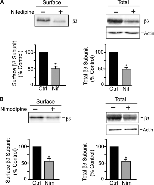
FIGURE 1.
Blockade of L-type VGCCs reduces expression of GABAARs. A, hippocampal neurons (18–21 DIV) were treated with 10 μm nifedipine (Nif) for 24 h and then biotinylated with Sulfo-NHS-biotin and lysed in RIPA buffer. Cell-surface proteins were isolated with immobilized avidin. Immunoblots show the total and cell-surface levels of GABAAR β3 subunits as indicated. Graphs represent quantification of band intensities of cell-surface and total β3 subunits normalized to controls (Ctrl). Data represent the mean ± S.E. percentage of control values. *, significantly different from the control (p < 0.01; t test; n = 4). B, nimodipine reduced the expression of the cell-surface and total levels of GABAAR β3 subunits. Hippocampal neurons were incubated with 15 μm nimodipine for 24 h and then biotinylated with Sulfo-NHS-biotin and lysed in RIPA buffer. Immunoblots show the cell-surface and total levels of GABAAR β3 subunits as indicated. Graphs represent quantification of band intensities, and data represent the mean ± S.E. percentage of control values. *, significantly different from the control (p < 0.05; t test; n = 3).

