FIGURE 6.
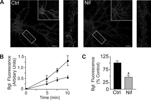
Insertion of GABAARs is modulated by Ca2+ influx through L-type VGCCs. A, images of pHluorin fluorescence and Alexa 594-conjugated α-Bgt staining at 5 min of insertion in the presence or absence of nifedipine (Nif; 10 μm) as indicated. Hippocampal neurons expressing BBSβ3 (18–21 DIV) were treated with or without nifedipine (10 μm) for 24 h. Neurons were then incubated with 10 μg/ml unlabeled α-Bgt to block existing cell-surface BBSβ3, washed, incubated for 5 min with 1 μg/ml Alexa 594-conjugated α-Bgt to label newly inserted BBSβ3, and fixed. The boxed areas in the upper right-hand corners show pHluorin fluorescence. The rectangles in the left panels (Alexa 594-conjugated α-Bgt staining) are enlarged in the right panels. B, increase in BBSβ3 insertion over time with or without nifedipine (10 μm). The assay was performed as described for A. C, graph showing quantification of Alexa 594-conjugated α-Bgt fluorescence intensity at 5 min of insertion for control (Ctrl) and nifedipine (10 μm)-treated neurons. Data represent the mean ± S.E. percentage of control BBSβ3 values. *, significantly different from the control (p < 0.01; t test; n = six to eight neurons from two independent cultures).

