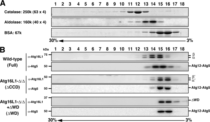
FIGURE 3.
Sucrose density gradient analysis of the Atg16L1 complex. A, standard proteins were separated using discontinuous sucrose density gradient centrifugation as described under “Experimental Procedures.” Fractions were subjected to SDS-PAGE and stained with Coomassie Brilliant Blue. B, cytosolic fractions of wild-type MEFs, Atg16L1-Δ/ΔMEFs expressing the empty vector, or Atg16L1-ΔWD were separated using discontinuous sucrose density gradient centrifugation as in A. Fractions were subjected to Western blotting using the indicated antibodies.

