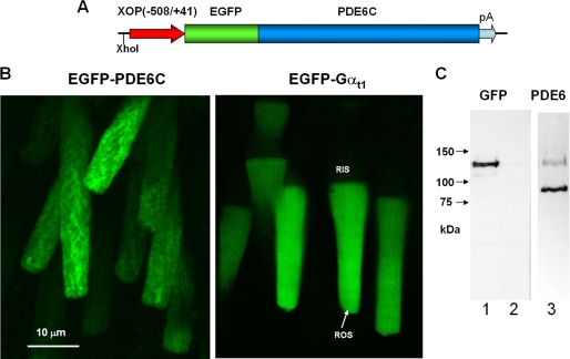
FIGURE 1.
Expression of EGFP-PDE6C in transgenic rods. A, map of the transgene. The XhoI site was used to linearize the pXOP(508/+41)EGFP-PDE6C plasmid for production of transgenic X. laevis embryos. B, EGFP fluorescence in living photoreceptor cells expressing EGFP-PDE6C and EGFP-Gαt. C, immunoblotting of retinal extracts from transgenic (lanes 1 and 3) and non-transgenic retinas (lane 2) with anti-GFP B-2 monoclonal antibody (Santa Cruz Biotechnology, Inc., Santa Cruz, CA) (lanes 1 and 2) and anti-PDE6 MOE antibodies (CytoSignal) (lane 3).

