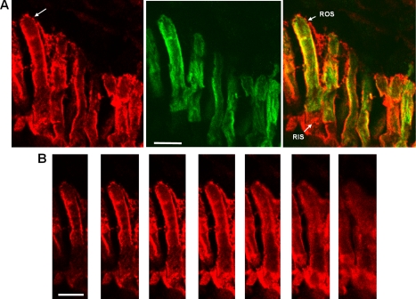
FIGURE 3.
Co-localization of EGFP-PDE6C with endogenous PDE6 in transgenic rods. A, red, immunofluorescence of a retinal cryosection of a light-adapted transgenic tadpole stained with anti-PDE6 MOE antibody; green, EGFP fluorescence; overlay. Bar, 10 μm. B, MOE antibody immunofluorescence. Z-Scans with an interval of 1 μm through the retina cryosection in A showing the rod cell identified by the arrow. Bar, 10 μm.

