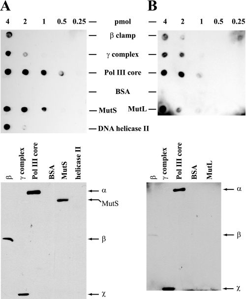
FIGURE 8.
Interactions of MutL and MutS with components of Pol III holoenzyme. β clamp, γ complex, Pol III core, BSA, MutS, MutL, and DNA helicase II were applied as indicated to nitrocellulose membranes (upper panels). Alternatively, proteins samples were subjected to SDS-PAGE on 10% gels (lower panels; 4-pmol sample load) followed by transfer to nitrocellulose membranes. The membranes were used for far Western analysis by incubation with MutL (A) or MutS (B), followed by immunochemical visualization of membrane-bound MutL and MutS (“Experimental Procedures”). SDS gel species that bind MutL and MutS are indicated to the right of each gel transfer, with identification based on parallel gels that were stained with Coomassie Blue. No signals were observed with otherwise identical membranes when MutS or MutL incubation steps were omitted (not shown).

