
Figure 1.
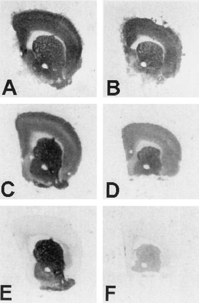
Receptor binding autoradiography of normal and R6/2 mice brains. Representative coronal sections of control (A, C, and E) and transgenic R6/2 mouse brain hemispheres (B, D, and F) subjected to [3H]AMPA binding (A and B), [3H]QNB binding for muscarinic acetylcholine receptors (C and D), or [3H]SCH-23390 binding for D1-like receptors (E and F). With all ligands, the R6/2 mice have lower levels of binding, but the extent of receptor binding decrease varies greatly, with D1 DARs most affected, mAChRs somewhat less affected, and AMPA subtype of ionotropic GluR only slightly decreased.