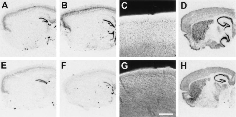
Figure 2.
In situ hybridization and immunofluorescence for mGluRs in control (A–D) and transgenic R6/2 (E–H) mice. In situ hybridization for mGluR2 mRNA in 4-week-old (A and E) and 12-week-old mice (B and F). mGluR2 mRNA hybridization signal appears dark on this film image. Note decreased labeling of cortical neurons in 12-week-old R6/2 mice; some decrease in cortical signal can be noticed in 4-week-old animals. (C and G): Fluorescent immunohistochemistry using an antibody that recognizes mGluR2 and mGluR3; signal appears bright in this image. Bar = 20 μm. Decreased mGluR2/3 immunoreactivity is apparent in the cortex of R6/2 mice. (D and H): In situ hybridization for mGluR5 mRNA.

