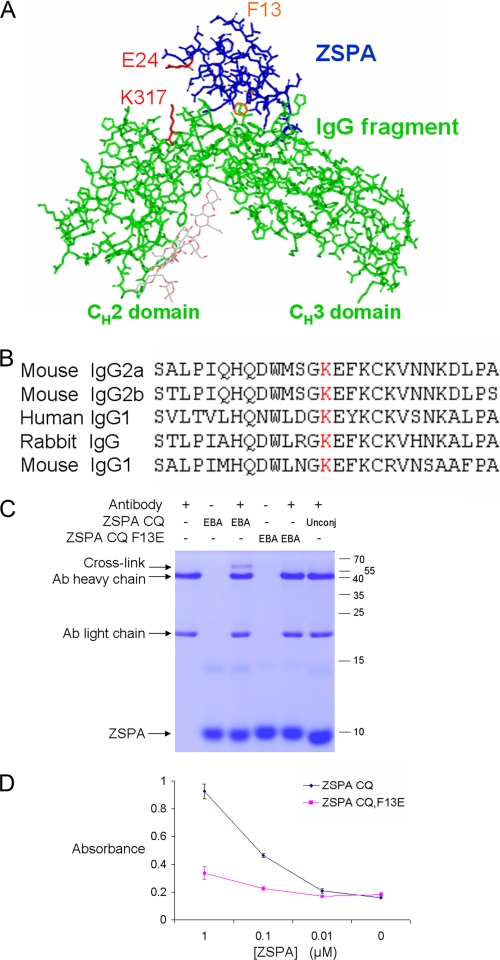
FIGURE 3.
Reaction of electrophilic ZSPA with immunoglobulin. A, design of electrophilic ZSPA. Guided by the Protein Data Bank structure 1FC2, ZSPA E24 (marked in red) was mutated to Cys, so that EBA attached here should be in proximity with the conserved lysine 317 (marked in red) on the antibody heavy chain. To block the non-covalent interaction, as a negative control, Phe-13 (marked in orange) was mutated to Glu. B, conservation of the target lysine (marked in red) in different antibody classes from various species. C, electrophilic ZSPA reaction with antibody. ZSPA CQ-EBA was incubated with antibody (Ab) and analyzed by SDS-PAGE and Coomassie staining. Negative controls were with unmodified ZSPA CQ (Unconj) or ZSPA CQ F13E. D, F13E mutation in ZSPA blocks binding to antibody. Plates were coated with ZSPA CQ or ZSPA CQ F13E and probed by ELISA with goat anti-mouse IgG-horseradish peroxidase. Means of triplicate measurements are shown ± 1 S.D.

