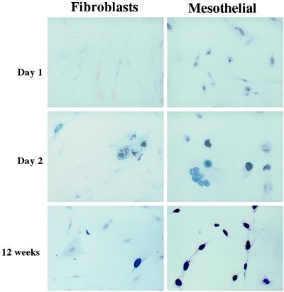
Figure 1.
Tag immunostaining. WI38 HF (Left) and HM3 (Right), at the indicated time after infection. Similar results were obtained with the other cells. No substantial differences were observed among HM1–3. In HM, the nuclear staining was punctate at 24 h, then it became granular, with the formation of intranuclear bodies, and finally obscured the entire nucleus. In HF, only a fraction of cells expressed Tag. These cells formed ill-looking cell clumps and giant cells, with clear evidence of cytopathic effects, such as vacuolization and lysis. (Original magnification, ×400.)

