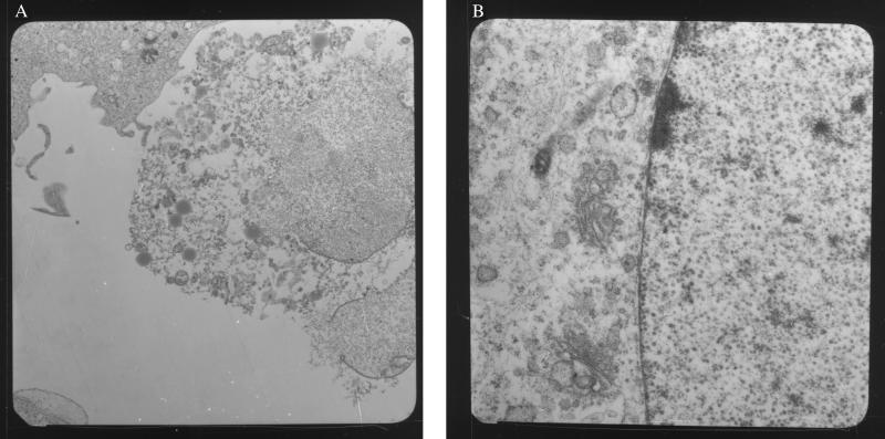
Figure 2.
EM of WI38 HF (A) and HM3 (B) infected with SV40 72 h earlier. Note that infected HF are lysed and full of viral particles (the individual viral particles are not clearly visible at this magnification). HM, instead, have intact nuclear membrane, and viral particles (round structures) are seen only in the nucleus (right side of the photograph). (Original magnifications: A, ×4, 400; B, ×20,000.) The same results were obtained when HM2 were used; HM1 were not tested.

