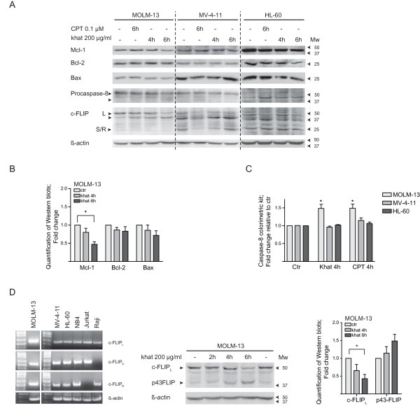
Figure 5.
Khat induced c-FLIP cleavage and Mcl-1 attenuation in khat sensitive MOLM-13. (A) MOLM-13, MV-4-11 and HL-60 were exposed to 200 μg/ml khat and 0.1 μM CPT for 4 and/or 6 hrs and evaluated for selected protein changes by Western blotting techniques. (B) Selected results for MOLM-13 were quantified using the KODAK Image Station 2000R software; the values normalized to the actin loading control and fold changes calculated. The asterisk indicates a significant reduction in Mcl-1 levels (p < 0.05). (C) MOLM-13, MV-4-11 and HL-60 were exposed to 200 μg/ml khat and 0.1 μM CPT for 4 hrs and caspase-8 activity evaluated using a Caspase-8 colorimetric kit. Fold change in enzymatic activity was determined by comparing the khat- and CPT-exposed samples to untreated controls. (D) Reverse transcription PCR was used to determine the expression of different c-FLIP RNA isoforms in the leukemic cell lines tested. Jurkat and Raji cells were utilized as controls since they are known to express the cFLIPS and c-FLIPR isoforms, respectively. β-Actin served as loading control. MOLM-13 was exposed to 200 μg/ml khat for 2, 4 and 6 hrs and evaluated for c-FLIPL cleavage by Western blotting techniques. The results were quantified, normalized to the actin loading control and fold changes calculated. The asterisk indicates a significant reduction in c-FLIPL levels (p < 0.05).

