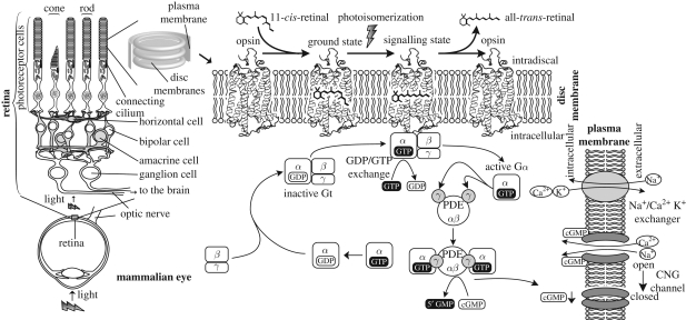
Figure 1.
A diagram showing the mechanism of phototransduction in mammalian eyes. Light is captured by two specialized morphologically distinct photoreceptor cells derived from neurons: rods and cones that have the same molecular mechanism. Opsins in these cells absorb photons and form a signalling state, which can bind to and activate the G protein by catalysing the exchange of GDP to GTP. The GTP-bound Gα dissociates from Gβγ exposing its active site. Activated Gα binds to its effector, PDE (cyclic nucleotide phosphodiesterase), and activates it. PDE breaks the phosphodiester bond of cGMP producing 5′GMP, and the decrease in the concentration of cGMP causes CNG (cyclic nucleotide gated) channels to close, which creates a hyperpolarization response in the photoreceptor cells. Light-activated rhodopsin is thermally unstable and the chromophore eventually detaches from the opsin. The hyperpolarization of the membrane potential of the photoreceptor cell modulates the release of neurotransmitters to downstream cells. The light signal is transmitted through different cells, finally reaching ganglion cells which form the optic nerve and project to the brain.

