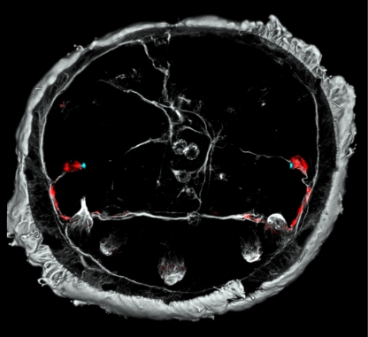
Figure 2.
The axonal scaffold of the larval brain of P. dumerilii at 48 hpf. Rhabdomeric PRCs (red) projecting to the ciliary girdle, where they locally alter the strength of ciliary beating (Jékely et al. 2008). Grey colour indicates axons and cilia labelled by an antibody directed against acetylated tubulin. Blue dots have been added following phalloidin stainings that specifically label the actin filaments of microvilli making up the rhabdomeres. Photograph courtesy of G. Jékely.

