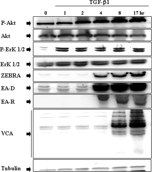
FIGURE 2.
Time course of TGF-β1-induced phosphorylation of Akt and ERK 1/2, and EBV lytic gene expression in Mutu-I cells. Mutu-I cells were incubated in the presence of TGF-β1 (2 ng/ml) for various periods of time. The cells were lysed, and equal amounts of proteins were separated by SDS-PAGE. Phosphorylated Akt and Akt were analyzed, respectively, with anti-phospho-Akt and Akt antibodies by Western blotting. The membrane was then reprobed separately with a panel of specific antibodies directed against phospho-ERK 1/2, ERK 1/2, ZEBRA, EA-D, EA-R, and VCA. The amounts of protein loaded were assayed by reprobing the membrane with anti-tubulin antibody.

