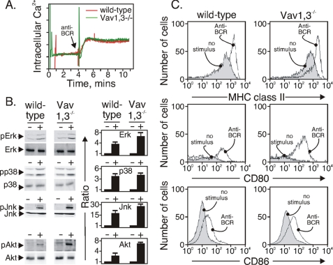
FIGURE 5.
Vav1,3 is not required for BCR-induced signaling events. A, anti-BCR-stimulated Ca2+ influx (arrow) of INDO-1-loaded B cells from the indicated strains. The data are representative of three separate experiments. B, anti-BCR-stimulated (+) activation of MAP kinase modules and pAkt in B cells from the indicated strains. Equal amounts of protein were loaded on the gel and quantitated as described under “Experimental Procedures.” The data are representative of four separate experiments. C, expression of MHC class II, CD80, and CD86 in unstimulated or anti-BCR-stimulated B cells from the indicated strains. Surface proteins were measured after 24 h with anti-BCR, and the data are representative of two separate experiments.

