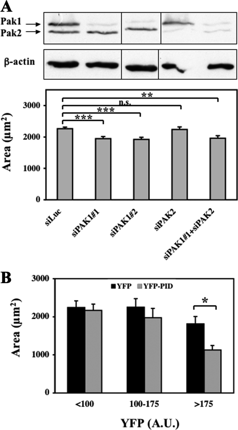
FIGURE 3.
Requirement of endogenous PAK1 activity for cell spreading. A, depletion of PAK1 inhibits cell spreading. HEK-HT cells were transfected with the indicated siRNAs. Three days later, they were trypsinized, re-plated on fibronectin-coated dishes, and fixed after 5 h of spreading. Cell areas were measured with ImageJ (n ≥ 75) and statistically analyzed. Depletion of PAK1 and PAK2 was >80% according to quantification by ImageJ. B, inhibition of PAK activity inhibits cell spreading. COS-7 cells expressing YFP or the YFP-PID fusion (PAK Inhibitory Domain) were trypsinized, re-plated on fibronectin-coated dishes and analyzed 4 h later. Area and average YFP fluorescence intensity were measured for each cell in two independent experiments (YFP cells, n = 65; YFP-PID cells, n = 101). Cells were divided into three groups according to their level of YFP fluorescence: <100, 100–175, and >175 (arbitrary units). The difference between the average area of YFP cells and YFP-PID cells is statistically significant for the third group of cells expressing the highest level of PID. Bars represent ± S.E. ***, p < 0.001; **, p < 0.005; *, p < 0.01, n.s. = not significant, according to Student's t test.

