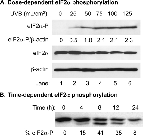
FIGURE 1.
Dose-dependent analysis of UVB-induced phosphorylation of eIF2α in HaCaT cells. A, Western blot analysis using cell extracts prepared from UVB-irradiated HaCaT cells. The amounts of total eIF2α, phosphorylated eIF2α, and β-actin were probed with corresponding antibodies. The levels of phosphorylated eIF2α were normalized by the levels of β-actin and expressed as a percentage of phosphorylated eIF2α. Lane 3, with an UVB dose of 50 mJ/cm2, was set to 1. The rest of the lanes were normalized accordingly. B, Western blot analysis using cell extracts prepared from UVB-irradiated and pETFVA−-eIF2α transfected COS-1 cells. The amounts of both eIF2α and phosphorylated eIF2α were probed with an antibody against eIF2α. The percentage of phosphorylated eIF2α was calculated as eIF2α-P/(eIF2α + eIF2α-P).

