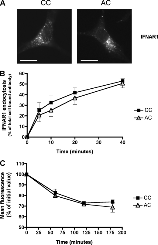
FIGURE 4.
IFNAR1 palmitoylation is not involved in IFNAR1 endocytosis and IFNAR1 half-life at the cell surface. A, endocytic pattern of the wild-type and AC IFNAR1 subunits. L929R2 cells stably transfected with wild-type IFNAR1 or the AC IFNAR1 mutant were processed for endocytosis as described in Fig. 1. Scale bar, 20 μm. B, FACS analysis of IFNAR1 endocytosis. L929R2 cells expressing either wild-type IFNAR1 or the IFNAR1 AC mutant were labeled with AA3 anti-IFNAR1 mAb prior endocytosis at 37 °C with 1000 units/ml IFN-α2b for the indicated times. Remaining cell surface IFNAR1-AA3 complexes were quantified by flow cytometry. Results are expressed as the disappearance of total cell-bound antibody from the cell surface. Each value is the mean ± S.D. of triplicate experiments. C, cell surface down-modulation of wild-type and AC IFNAR1 subunits. The cell surface expression of IFNAR1 was measured by flow cytometry in L929R2 cells expressing either wild-type or AC IFNAR1 subunits in the presence of cycloheximide and 1000 units/ml IFN-α2b for the indicated times.

