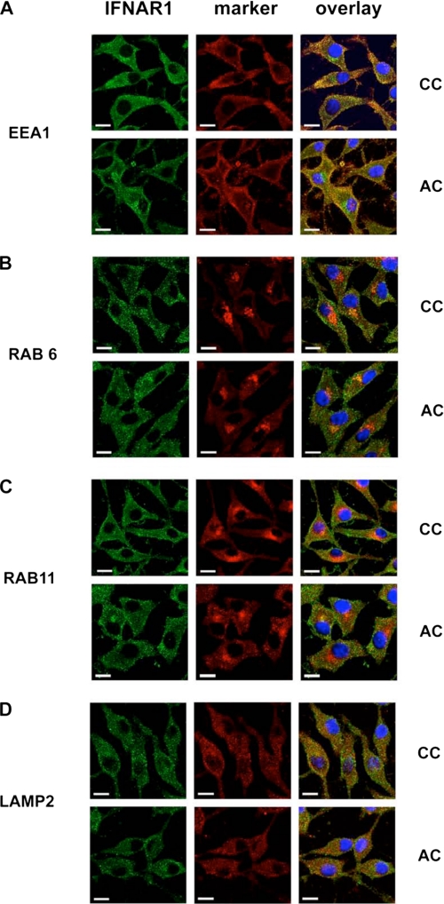
FIGURE 5.
Similar intracellular distribution of wild-type IFNAR1 and palmitoylation-deficient AC IFNAR1 mutant. L929R2 cells stably transfected with wild-type IFNAR1 or the AC mutant were fixed, permeabilized, and co-labeled with anti-IFNAR1 34F10 mAb and antibodies directed either against the early endosome marker EEA1 (A), the Golgi apparatus marker Rab6 (B), the recycling endosome marker Rab11 (C), or the lysosomal marker Lamp2 (D). Secondary antibodies coupled with Alexa 488 (green) and with Cy3 (red) were used to reveal IFNAR1 and cellular markers, respectively. 4′,6-Diamidino-2-phenylindole was added to detect nuclei. Cells were imaged with a confocal Leica microscope. Scale bar, 15 μm.

