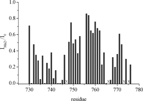
FIGURE 9.
Manganese binding to polycystin-2-(680–796). To a sample of 500 μm unlabeled polycystin-2-(680–796) in 5 mm Tris-HCl, pH 6.8, 100 mm NaCl, 2 mm dithioerythritol, 0.1 mm DSS, 5 mm CaCl2 in 90% H2O, 10% D2O a concentrated solution of MnCl2 was added, and 1H-1H NOESY spectra were recorded. The ratio of the cross-peak volumes in the absence (I0) and the presence (IMn2+) of MnCl2 was determined. For every residue the intra-residual cross-peak with the lowest ratio of IMn2+/I0 at a MnCl2 concentration of 0.4 mm was plotted as a function of the sequence. Residues where intra-residual cross-peaks are too weak to be observed in the absence of MnCl2 are labeled by x.

