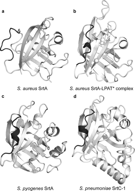
FIGURE 6.
Some sortase enzymes contain a preformed binding pocket for the LPXTG sorting signal. The figure compares the three-dimensional structures of sortase enzymes that recognize LPXTG sorting signals. Four structures of SrtA-type sortases are shown. a, the crystal structure of S. aureus apo-SrtAΔN59 (Protein Data Bank code 1t2p) (31); b, the NMR structure of the S. aureus SrtAΔN59-LPAT* complex; c, the crystal structure of S. pyogenes apo-SrtAΔN81 (Protein Data Bank code 3fn5) (37); d, the crystal structure of S. pneumoniae apo-SrtC-1ΔN16 (Protein Data Bank code 2w1j) enzyme (38). The β6/β7 loop in each structure is highlighted in dark gray to emphasize differences and similarities. The comparison reveals that the β6/β7 loops in the S. pneumoniae and S. pyogenes enzymes adopt a closed helical conformation similar to the substrate-bound form of the S. aureus SrtA enzyme in the SrtAΔN59-LPAT* complex. This is in marked contrast to the S. aureus apoenzyme, which adopts an open conformation. Note that the structure of S. pneumoniae apo-SrtC-3ΔN31 has also been determined and is not shown here, because it is generally similar to the structure of S. pneumoniae apo-SrtC-1ΔN16 (38).

