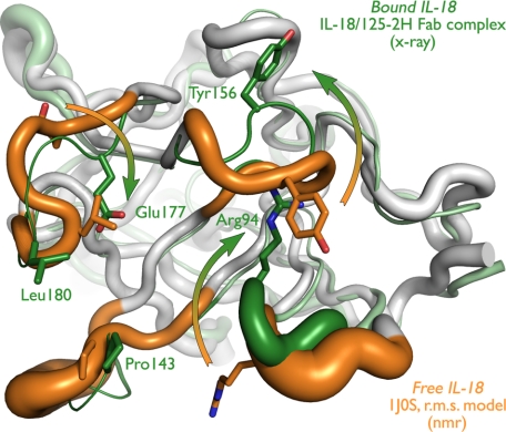
FIGURE 1.
Free and 125-2H-bound IL-18 exhibit large conformational differences. Free (white and orange; NMR (Protein Data Bank entry 1J0S)) and 125-2H-bound (gray and green; x-ray (this work)) IL-18 are overlaid. In these schematic diagrams, the tube width represents the (equivalent) temperature factor of the corresponding Cα atom (equivalent temperature factor Bequiv = 8π2〈u2〉/3, where 〈u2〉 is the r.m.s. deviation of the 20 NMR models). The four IL-18 loops that bind to 125-2H are colored orange (free) or green (bound). Note the significant movement (arrows) of Arg94, Tyr156, Glu177, and Leu180, and associated surface loops, which far exceeds the observed positional variability (tube diameter) in the structures. See also supplemental Fig. 1, a and b.

