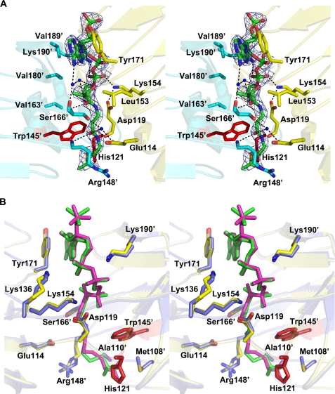
FIGURE 3.
A, stereoview of the donor-binding site with electron density of acetyl-CoA in a refined 2Fo − Fc map contoured at 1.5σ. Bound acetyl-CoA molecule is shown using green for carbon and phosphorus atoms, with other atoms colored according to atom type (nitrogen, blue; oxygen, red; and sulfur, gold). Interacting residues from one monomer are shown in yellow, and interacting residues from the adjoining monomer are shown in cyan. Water molecules are depicted as blue spheres, and His-121 and Trp-145′ are displayed as red sticks. Polar contacts are shown as dotted lines. B, stereoview of a structural alignment between the complexes of OatWY with acetyl-CoA and a nonhydrolyzable CoA analog. Key residues that interact with the donor substrate are represented in yellow for the acetyl-CoA complex and in blue for the S-(2-oxopropyl)-CoA complex. Acetyl-CoA and S-(2-oxopropyl)-CoA molecules are depicted in green and magenta, respectively. His-121 and Trp-145′ are represented in red.

