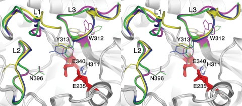
Fig. 2.
Active site of velaglucerase alfa. Stereo representation of an overlay of the active sites of imiglucerase (blue and magenta) and velaglucerase alfa (yellow and green). Catalytic residues are shown as red sticks. Loops near the entrance to the active site are indicated (L1, loop 1; L2, loop 2; L3, loop 3).

