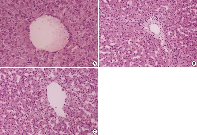
Fig. 5.
(A) Hematoxylin-eosin-stained specimen before clamping the portal triad in pigs (×100). Image shows normal portal tract in the pig liver. (B) Specimen after 4 hr since the first clamping in the non-ligation group (×100). Widened portal spaces with infiltration of a few inflammatory cells and ischemic changes of hepatocytes in porto-to portal space are shown but there are no necrotic hepatocytes. The sinusoidal spaces are dilated. (C) Specimen after 20 hr since the first clamping in the non-ligation group (×100). There are no histologic differences from those of (B).

