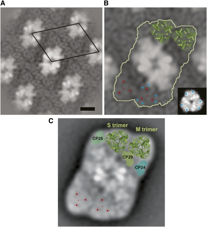
Figure 3.
Photosystem II Supercomplex Structure in Wild-Type and koLhcb3 Plants.
(A) Projection map of semicrystalline wild-type Arabidopsis PSII-LHCII membranes with the repeating unit (unit cell) indicated by the black diamond. Bar = 10 nm.
(B) koLhcb3 line N520342 crystal map showing the C2S2M2 supercomplex particle (white outline) and the position of the S- and M-trimers from the high-resolution x-ray map of LHCII is superimposed on the top trimers (green). The small red dots mark the position of the S- and M-trimers, and the larger red dots surrounding these show positions of high contrast. Blue dots indicate two recognizable densities at the periphery. Inset: A LHCII trimer with comparable densities in EM and x-ray maps indicated with blue dots.
(C) Projection map of the wild-type C2S2M2 supercomplex obtained from single particle averaging for comparison (Kouril et al., 2005). Positions within the S- and M-trimers compatible to the crystal two-dimensional map are indicated by red dots.

