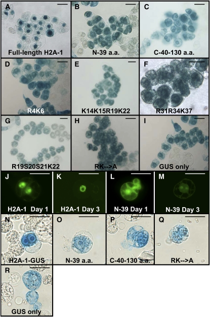
Figure 5.
Subcellular Localization of Full-Length Histone H2A-1 and Various Mutant Peptides Fused to Either GUS or YFP Reporter Proteins.
(A) to (I) X-gluc staining of stably transformed tobacco BY-2 cell lines expressing the indicated GUS fusion proteins. Note that expression of an unfused gusA gene results in cytoplasmic staining of the cells (I), whereas expression of a full-length H2A-1-GUS fusion protein results in predominantly nuclear staining (A). Expression of the various H2A-1 mutant peptides results in predominantly cytoplasmic, especially perinuclear, staining ([B] to [H]).
(J) to (M) Epifluorescence images of tobacco BY-2 protoplasts 1 d ([J] and [L]) or 3 d ([K] or [M]) after transfection with the indicated constructions. Note that the full-length H2A-1-YFP fusion protein localizes predominantly to the nucleus ([J] and [K]) but that the N-terminal H2A-1-YFP fusion protein localizes throughout the cell ([L] and [M]). YFP fluorescence is shown in yellow-green.
(N) to (R) X-gluc staining of transfected tobacco BY-2 protoplasts expressing the indicated GUS fusion proteins 1 d after transfection.
Bars = 50 μM.

