Figure 6.
PIN Expression and Localization in the Wild Type and nov-1.
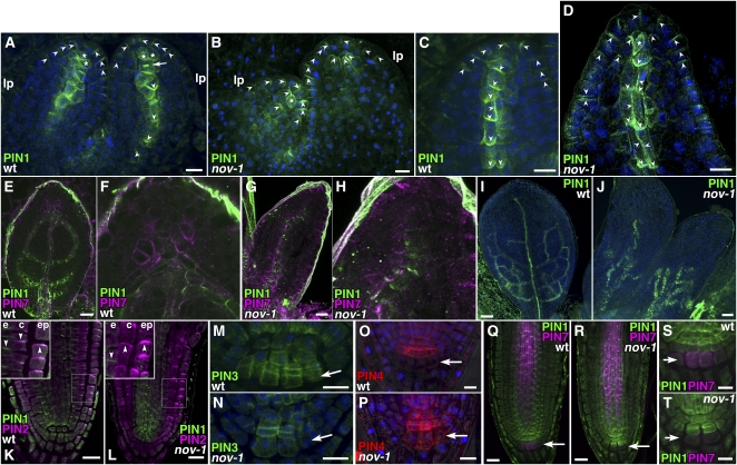
(A) to (J) Immunostaining of PIN1 and PIN7 in leaf primordia. PIN1 (green in [A] to [J]), PIN7 (magenta in [E] to [H]) and DAPI (blue in [A] to [D], [I], and [J]). Images were obtained from the first two leaf primordia of the wild type ([A], [C], [E], [F], and [I]) and nov-1 ([B], [D], [G], [H], and [J]). Asterisks indicate cells with nonpolar PIN1. An arrow in (A) indicates PIN1 accumulation at a transverse cell division plane. lp, leaf primordium.
(K) and (L) Immunostaining of PIN1 and PIN2 in the root tip. Double staining of PIN1 (green) and PIN2 (magenta) in root tips of the wild type (K) and nov-1 (L). Insets show boxed areas enlarged. e, endodermis; c, cortex; epi, epidermis.
(M) and (N) Immunostaining of PIN3 in the root tip. PIN3 (green) and DAPI (blue) in the root stem cell niche of the wild type (M) and nov-1 (N).
(O) and (P) Immunostaining of PIN4 in the root tip. PIN4 (red) and DAPI (blue) in the root stem cell niche of the wild type (O) and nov-1 (P).
(Q) to (T) Immunostaining of PIN1 and PIN7 in the root tip. Double staining of PIN1 (green) and PIN7 (magenta) in root tips of the wild type ([Q] and [S]) and nov-1 ([R] and [T]). Arrowheads indicate the polarity of PIN proteins for clarity. Arrows in (M) to (T) indicate the tier three of columella root cap cells.
Bars = 10 μm in (A) to (D), (M) to (P), (S), and (T), 20 μm in (E), (G), (K), (L), (Q), and (R), and 50 μm in (I) and (J).

