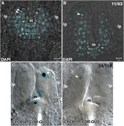
Figure 7.
DAPI Staining and CYCB1pro:CDB:GUS Expression in Wild-Type Leaf Primordia.
(A) and (B) Wild-type leaf primordia were stained with DAPI. Arrowheads indicate transversely aligned condensed nuclear DNA at the apical ends of provascular cells for the midvein.
(C) and (D) The wild type containing CYCB1pro:CDB:GUS was examined for GUS activity. Arrowheads indicate GUS expression at the apical ends of provascular cells for the midvein. All images were taken from the lateral view of leaf primordia. Shown in the top left corner of (B) and (D) are fractions of primordia with displayed features, respectively.
lp, leaf primordium; m, shoot apical meristem. Bars = 10 μm.

