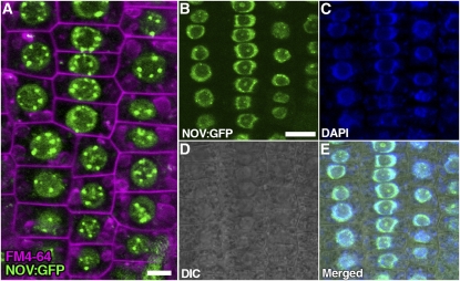
Figure 9.
Nuclear Localization of NOV:GFP.
(A) to (E) Subcellular localization of NOV:GFP was examined in root epidermal cells of seedlings containing NOVpro:NOV:GFP (NOV:GFP). Shown in (A) is a confocal image of root epidermal cells for NOV:GFP (green) and FM4-64 staining (magenta, plasma and endocytic membranes). NOV:GFP (green in [B]), DAPI (blue in [C]), differential interference contrast (DIC) optic image (D), and merged image (E) are also shown. NOV:GFP is specifically localized in the nucleus. Bars = 5 μm in (A) and 10 μm in (B) (equal scale in [B] to [E]).

