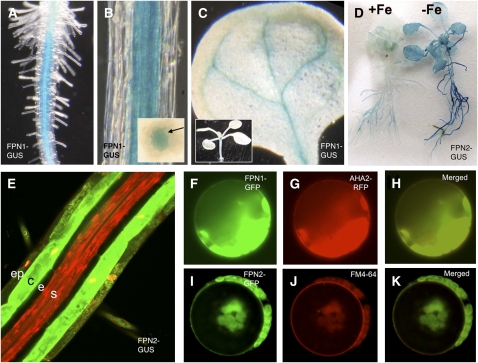
Figure 2.
Localization of FPN1 and FPN2.
Eleven-day-old FPN1-GUS plants show staining in the stele ([A] and [B]), root-shoot junction, and veins of the cotyledons (C). Inset in (B) shows staining in stele of root cross section. Inset in (C) shows staining in the seedling root-shoot junction. When 2-week-old FPN2-GUS plants are transferred from B5 to –Fe minimal medium for 3 d, GUS staining is very dark in the root and is also present in the shoot (D). Staining with propidium iodide (red) and the fluorescent GUS substrate, ImaGene Green, shows FPN2-GUS expression primarily in the cortex but also in the epidermis and root hairs (E). When FPN1 and FPN2 are fused to GFP and transiently expressed in protoplasts, FPN1-GFP localizes to the plasma membrane (F), as does the plasma membrane marker AHA2-RFP (G); FPN2-GFP localizes to the vacuole (I), as does the vacuole marker dye FM4-64 (J). Overlays of GFP and the markers are shown in (H) and (K).

