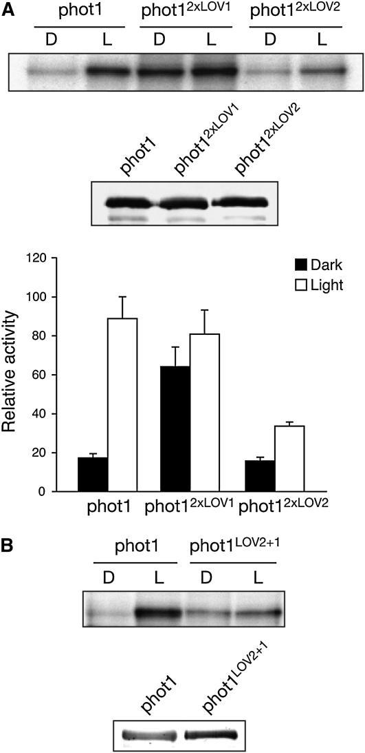
Figure 4.
Autophosphorylation Activity of Full-Length phot1 Domain-Swap Proteins Expressed in Insect Cells.
(A) Autoradiograph showing light-dependent autophosphorylation of wild-type phot1, phot12xLOV1, and phot12xLOV2 in protein extracts isolated from insects cells. Samples were given a mock irradiation (D) or irradiated with white light (L) at a total fluence of 10,000 μmol m−2 prior to the addition of radiolabeled ATP. Immunoblot analysis of phot1 protein levels is shown below. Kinase activity was quantified by phosphor imaging and expressed as a percentage of maximal phosphorylation activity. Standard errors are shown (n = 3).
(B) Autophosphorylation activity of the phot1LOV2+1 domain-swap protein relative to wild-type phot1. Immunoblot analysis of phot1 protein levels is shown below.

