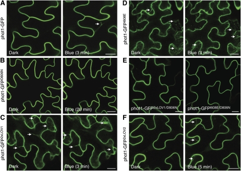
Figure 7.
Subcellular Localization of phot1-GFP and Domain-Swap Proteins in N. benthamiana.
(A) Fluorescence images showing the rate of phot1-GFP internalization in tobacco leaf epidermal cells at 3 min after a 30-s irradiation with blue light. The white arrow indicates internalization.
(B) Inactivation of phot1 kinase activity (phot1-GFPD806N) inhibits blue light–dependent internalization.
(C) phot1-GFP2xLOV1 shows internalization in the absence of a blue light stimulus. The white arrows indicate internalization.
(D) phot1-GFPI608E also shows constitutive internalization as indicated by the white arrows.
(E) Inactivation of phot1 kinase activity by incorporation of the D806N mutation blocks internalization of phot1-GFP2xLOV1 and phot1-GFPI608E in the absence of light.
(F) phot1-GFP2xLOV2 undergoes blue light–dependent internalization, similar to phot1-GFP.
Bars = 20 μm.
[See online article for color version of this figure.]

