Figure 1.
The U. maydis PMT Family.
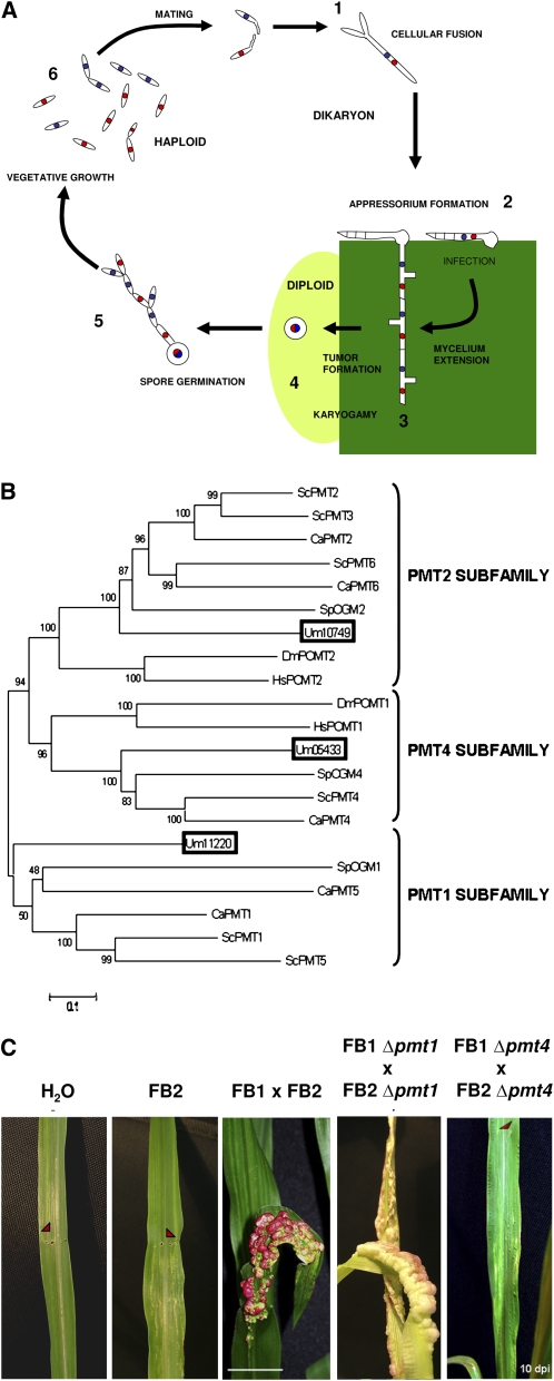
(A) Life cycle of U. maydis. Developmental stages of the U. maydis life cycle: 1, fusion of two haploid cells; 2, dikaryon filament invades plant cells to form the appressorium; 3, U. maydis proliferates and differentiates within tumor tissue; 4, the fungus produces diploid spores; 5, spores undergo meiosis; 6, U. maydis enters its haploid phase.
(B) Dendrogram of PMT proteins. PMT proteins of S. cerevisiae (ScPMT), S. pombe (SpOGM), C. albicans (CaPMT), D. melanogaster (DmPOMT), and H. sapiens (HsPOMT) were compared with the Um11220, Um10749, and Um05433 proteins (boxed). The black bar represents an evolutionary distance of 0.1 amino acid substitutions per site. Bootstrap support values are given at branch nodes. The phylogenetic analysis showed that a single putative U. maydis PMT protein was assigned to each of the three PMT subfamilies. A text file of the alignment used to generate this dendogram is available as Supplemental Data Set 1 online.
(C) Effects of pmt4 deletion on pathogenicity. Seven-day-old maize seedlings were inoculated with water, FB2, a cross of FB1 and FB2 wild-type strains, FB1 Δpmt1 and FB2 Δpmt1, or FB1 Δpmt4 and FB2 Δpmt4. Arrows point to the injection punctures. Ten days after infection (dpi), anthocyanin production and tumor formation were observed on plants infected with the wild-type and Δpmt1 crosses, while no infection symptoms were detected on plants inoculated with the pmt4 mutant strains. Bar = 2 cm.

