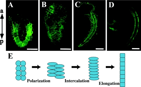
Figure 1. Convergent extension movement of ascidian notochord.
Confocal images of frontal section of H. roretzi stained with Alexa Fluor® 488 phalloidin to visualize cellular boundaries. (A) At the initial tail-bud stage, 40 post-mitotic notochord cells are aligned in two bilateral lines. Anterior is at the top. (B) Each notochord cell is polarized, elongates along the left–right axis and starts mediolateral intercalation. (C) The notochord is made of disc-shaped cells aligned in a single line after intercalation. (D) At the mid tail-bud stage, each notochord cell elongates in a process of post-convergent extension elongation. In consequence, the tail further elongates. (E) Representation of notochord morphogenesis. a, anterior; p, posterior. Scale bar, 100 μm.

