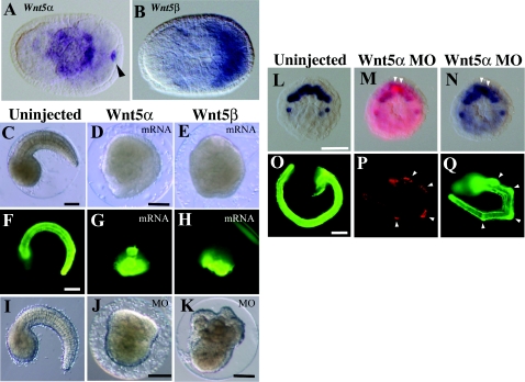
Figure 2. Effects of Wnt5 MOs and mRNAs injected into eggs.
(A, B) Expression of Hr-Wnt5α and Hr-Wnt5β genes at the neurula stage in developing notochord and muscle cells respectively. Anterior is to the left. Arrowhead indicates Hr-Wnt5α expression in the posterior pole, which is concentrated Hr-Wnt5α maternal mRNA, as it is a member of postplasmic/PEM RNAs in ascidians (Sasakura et al. 1998). (C–E) Morphology of uninjected and Wnt5α and Wnt5β mRNA-injected embryos at the tail-bud stage. (F–H) Detection of the notochord differentiation marker antigen, Not-1, in uninjected and Wnt5α and Wnt5β mRNA-injected embryos. (I–K) Morphology of uninjected, Wnt5α and Wnt5β MO-injected embryos at the tail-bud stage respectively. (L) Expression of Bra gene in ten notochord precursor blastomeres at the 110-cell stage. (M) Embryo co-injected with Wnt5α MO and a lineage tracer (rhodamine dextran) into a single notochord precursor (A7.3) blastomere of the 64-cell embryo. At the 110-cell stage, two sister blastomeres are labelled with red fluorescence (white arrowheads). (N) The same embryo normally expressed the Bra gene in notochord precursors, including the labelled cells. (O) The Not-1 antigen is expressed in notochord. The embryo was slightly overstained with the antibody, and the signal is spread in the trunk region. (P) At the tail-bud stage, several descendants of the injected blastomere can be recognized from their content of red fluorescent label. (Q) The same embryo expressed Not-1 antigen in notochord cells including the injected and labelled cells (arrowheads). Scale bar, 100 μm.

