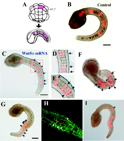
Figure 4. Effects of Wnt5α mRNA injected into a notochord precursor blastomere.
(A) Lineage illustration of notochord. (B) Tail-bud embryos co-injected with the lineage tracer and control FS Wnt5α mRNA into the A7.3 blastomere. (C) Injection of Wnt5α mRNA. Arrows indicate normally intercalated notochord cells. Arrowheads show cells that failed to intercalate. (D) Closer view of the blue rectangle in (C). Three notochord cells successfully intercalated. (E) Closer view of the red rectangle in (C). The two cells marked with arrowheads failed to intercalate with each other. (F–I) Four examples of Wnt5α mRNA-injected embryos to show the wide spectrum of abnormality in intercalation. In (I), post-convergent elongation of notochord cells is taking place. Scale bar, 100 μm.

