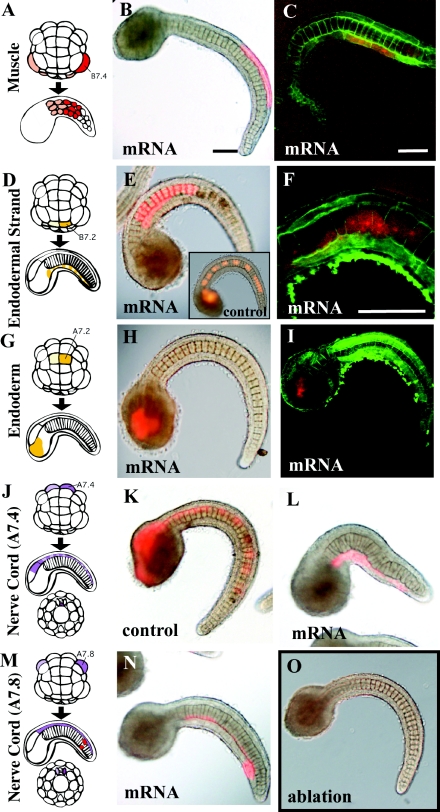
Figure 5. Effects of Wnt5α mRNA injected into various tissue precursors.
(A, D, G, J, M) Lineage illustration of muscle, endodermal strand, endoderm and nerve cord (A7.4 and A7.8) respectively (Nishida, 1987). Injected blastomeres and their descendants are indicated in dark colours. (B, E, H, K, L, N) Bright-field images are merged with fluorescent images to show the position of labelled descendant cells. (C, F, I) Confocal views. Cell boundaries are demarcated by green fluorescence. (B, C) The muscle precursor was injected. (E, F) The precursor of the endodermal strand was injected. The notochord is normal, but intercalation of the endodermal strand, which is present on this side of the notochord, was prevented as the labelled cells did not intercalate with the cells of the other bilateral side as they formed a continuous line. (F) is a confocal closer view of such a specimen. (E, insert) Control embryo. The endodermal strand is intermittently labelled in the tail. (H, I) The endoderm precursor was injected. (K, L) Control mRNA and Wnt5α mRNA were injected into the precursor of the ventral row (floor plate) of the nerve cord respectively. In the control, the nerve cord is intermittently labelled in the tail, indicating intercalation of left and right descendants. In Wnt5α mRNA-injected embryos, the labelled cells failed to intercalate as they formed a continuous line. (N) Wnt5α mRNA was injected into the precursor of the lateral row of the nerve cord in addition to two muscle cells at the tip of the tail (red). (O) Every A-line nerve cord precursor blastomere was ablated at the 64-cell stage by bursting them with injected seawater. The notochord of the embryo developed normally, indicating that the neural tube is dispensable for notochord morphogenesis. Scale bar, 100 μm.

