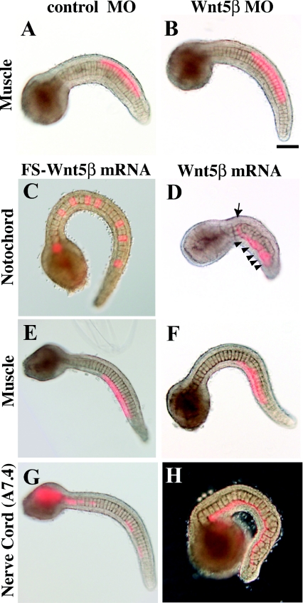
Figure 6. Effects of Wnt5β MO and mRNA injected into various tissue precursors.
(A, B) Control and Wnt5β MO were injected into the muscle precursor (B7.4) blastomere as Wnt5β is expressed in muscle. (C–H) Control FS Wnt5β mRNA and wild-type Wnt5β mRNA were injected into the precursors of notochord (A7.3), muscle (B7.4) and the ventral row of nerve cord (A7.4). Arrow indicates a normally intercalated notochord cell. Arrowheads show cells that failed to intercalate. (G) In the control, the nerve cord is intermittently labelled in the tail, indicating intercalation of left and right descendants. (H) In Wnt5β mRNA-injected embryos, the labelled cells failed to intercalate as they formed a continuous line. Scale bar, 100 μm.

