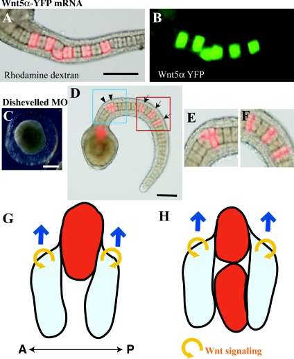
Figure 8. Wnt5α may function in a cell-autonomous manner.
(A, B) The lineage tracer (rhodamine dextran; red) and mRNA encoding the Wnt5α–YFP fusion protein (green) were co-injected into a notochord precursor blastomere. The green signal remains close to the injected descendant cells. (C) An embryo in which Dsh MO was injected into fertilized eggs. (D) An embryo in which Dsh MO was injected into a notochord precursor blastomere. Arrows indicate normally intercalated notochord cells. Arrowheads show cells that failed to intercalate. (E, F) Closer views of the blue and red rectangles shown in (D). Scale bar, 100 μm. (G) A descendant cell of the MO- and mRNA-injected blastomere (red) is present in isolation. Flanking normal cells (light blue) may migrate on the surface of the anomalous cell. Blue arrows indicate the direction of movement of the normal cells. Yellow circular arrows refer to the autocrine Wnt5α action. Anterior is to the left. A, anterior; P, posterior. (H) When anomalous cells are closely adjacent they fail to migrate on each other. See the text for further details.

