
Figure 4.
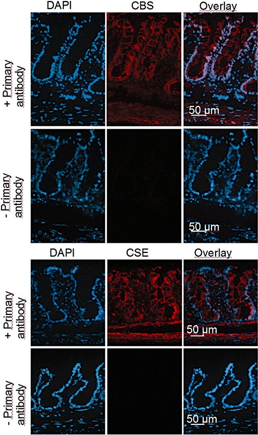
Distribution of cystathionine-β-synthase (CBS) and cystathionine-γ-lyase (CSE) immunoreactivity in rat colonic wall. The orientation of each picture is: lower part: muscularis propria; upper part: surface region of the colonic epithelium. Left column: nuclear staining with 4′,6-diamidino-2-phenylindoldilactate (DAPI) (blue); middle column: CBS signal (first row; Cy3-labelled, red) or CSE signal (third row; Cy3-labelled, red); right column: overlay of both. Second and fourth row: negative controls obtained in the absence of primary antibody against the respective enzyme. Typical results from three independent experiments.